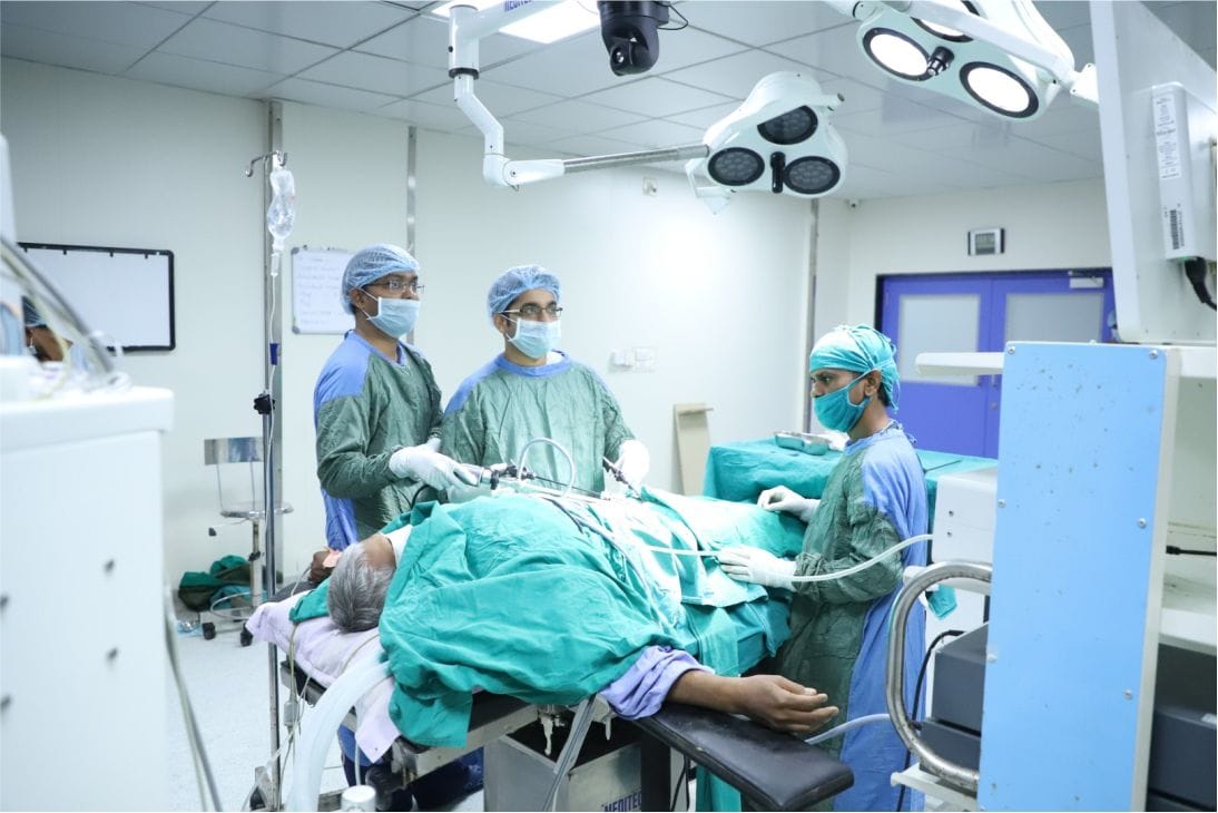
The surgery was carried out by the expert Oncosurgical team of Parul Sevashram Hospital without any iatrogenic injuries and minimal blood loss.
In a very challenging and interesting case recently, a 27 years old female patient from Madhya Pradesh presented to the Surgical Oncology department of Parul Sevashram Hospital with a mass occupying almost 2/3rd of the abdomen. The patient had amenorrhoea before 4 months with a rapidly increasing abdominal mass. She was diagnosed as a case of multiple pregnancies (Pregnancy test positive). She had a miscarriage after 2 months, with a persistent mass that was increasing in size. She had consulted earlier at multiple clinics and hospitals and was in distress due to the pain and pressure effects caused by the massive lump. Upon investigation at PSH in the form of a CT scan and MRI, she was diagnosed to have a huge mass in the left lumbar region involving the left kidney and adrenal gland. It was adherent to all the surrounding critical structures like the pancreas, spleen, left colon, stomach, duodenojejunal flexure, aorta, and left diaphragm. Hence without wasting any more time, the surgeons planned an exploratory laparotomy for the removal of the mass. The patient and relatives were explained that one or more of the structures adherent to the mass may have to be removed along with the mass, which may cause some postoperative morbidities.
“During the surgery, we were alarmed to see a huge mass of more than 20 cm filling the entire left side of the abdomen”, said Dr. Dipayan Nandy, OncoSurgeon, Parul Sevashram Hospital.
The left colon and its mesentery densely adhered to the mass, which was carefully separated without any injury to the blood supply of the colon, thus preventing bowel resection. The mass was found to be originating from the upper pole of the left kidney and involving the left adrenal gland. The spleen and its vessels were also adherents to the mass, which were meticulously dissected free, thus preventing splenectomy. The tail of the pancreas, posterior wall of the stomach, and the duodenojejunal flexure were all dissected carefully without any iatrogenic injuries, he added.
The mass was then dissected from the aorta and the renal vessels were identified and ligated.
The mass was densely adherent to the left diaphragm and hence wide excision of part of the left diaphragm was done en-bloc with the mass. The diaphragmatic defect was repaired primarily after placing an ICD in the left thorax.
The entire mass was then mobilized free from the retroperitoneum and radical left nephrectomy was done without any rupture or spillage. The mass was sent for the Histopathological examination, and after a thorough evaluation by the expert Histopathologists, it was established to be Wilm’s tumor.
There was a very minimal loss in the entire surgery and it was uneventful during the entire course of the surgery.
The patient got immediate relief from the pain and pressure effects of the mass and could start eating and moving around from 3rd post-op day.
“The combination of surgery by expert surgeons aided with cutting edge technology and perioperative management in our state-of-the-art ICU serves as a boon in treating such challenging cases”, said Dr. Geetika Madan Patel, Medical Director, Parul Sevashram Hospital.
Renal masses are detected late unless the patient has hematuria (blood in urine) or when it grows big enough to cause pain and other symptoms. Any suspicion of a renal mass should be investigated at the earliest since small renal tumors can be treated with partial nephrectomy. This helps to preserve more functional renal tissue and the patient does not have to live with the hazards of having a single kidney.


